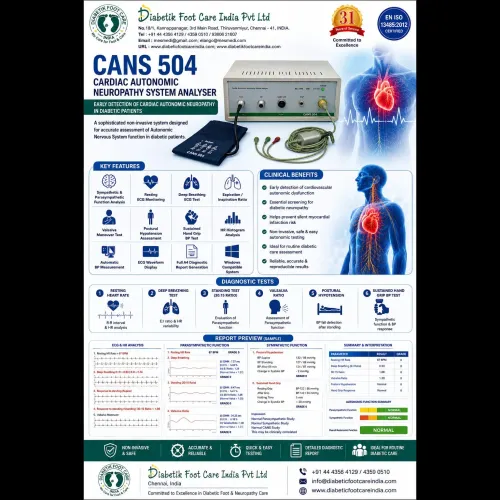
Vascular Screening Devices:
Diabetik Foot Care India Pvt Ltd is a pioneer in the manufacturing and distribution of medical devices for the management of diabetic foot and its complications.
Our products are working in as many as 34 countries. More than 25 field sales cum service engineers are working across India to take care of sales and after sale services. Most of the leading institutions treating diabetes are our clients.
Whenever one suspects Peripheral Arterial Disease (PAD), the clinician must perform few non-invasive vascular testing methods that are commercially available and widely implemented. They include the ankle brachial index ABI), the toe-brachial index(TBI), segmental Pressure Study and pulse volume recording(PVR), transcutaneous oxygen monitoring(TCPO2) and skin perfusion pressure(SPP).
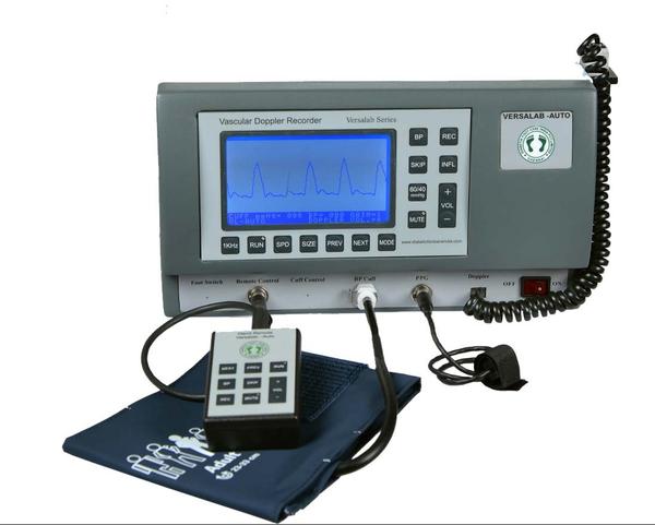
Ankle Brachial Index:
The ankle brachial index is the most well-known, non-invasive vascular testing tool. ABI test is performed with a Doppler and a blood pressure cuff. One calculates the ABI by dividing the ankle pressure by the brachial systolic pressure. An ABI of < 0.9 is abnormal and ABI values have a linear correlation with wound healing potential in lower extremity wounds. Patients with DM may have calcified and hardened lower extremity arterial walls that cannot be readily compressed and occluded with blood pressure cuffs. This produces falsely elevated ankle pressure readings that are often in the “normal ABI range” (0.9 to 1.3) or sometimes in the non-physiological range of above 1.3. However, Calcified leg arteries in Diabetes Mellitus or dialysis patients may yield falsely elevated ABI results.
Toe-Brachial Index:
The digital arteries in great toes are considered to be less affected by medial arterial calcification. One would calculate TBI by dividing the blood pressure of the great toe by the systolic brachial blood pressure. Toe pressure of > 50mmHg is considered normal. Toe pressure < 30 mmHg is considered severely ischemic. Toe Brachial Index (TBI) of less than 0.7 is considered abnormal.
Segmental Limb Pressures:
Once the ABI has been performed, segmental limb pressure measurement can aid in localizing stenosis or occlusions. Limb pressure cuffs are placed on the thigh, below knee, ankle and digit. The pressure at each segment is measured. A difference in pressure of >30 mmHg between the cuff sites suggests a significant arterial stenosis or occlusion present between the site.
Pulse-Volume Recording:
Pulse-volume recordings (PVRs) are plethysmographic tracings that detect changes in the volume of blood flowing through a limb. Using equipment similar to the segmental limb pressure technique, pressure cuffs are inflated to 65 mm Hg, and a plethysmographic tracing is recorded at various levels. A normal PVR is similar to a normal arterial pulse wave tracing and consists of a rapid systolic upstroke and a rapid down stroke with a prominent dicrotic notch. With increasing severity of PAD, the waveforms become more attenuated with a wide down slope and, ultimately, virtually absent waveforms.
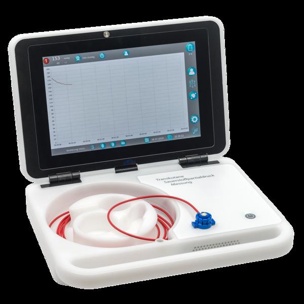
Transcutaneous Oxygen Monitoring - TCPO2:
Transcutaneous oxygen monitoring (TCOM) and skin perfusion pressure (SPP) are valuable tools in the wound care setting. As these methods are unaffected by calcified arteries and a higher pressure reading clinically correlates with increased wound healing potential. In addition, both devices allow strategic sensor placement in various locations around foot and ankle wounds. When combined these tools provide specific information relative to leg ischemia, wound healing potential, optimal amputation level and incision site determination for the lower extremities. Normal values are > 50 mmHg. Wound healing potential drops as TcPO2 values decline. Traditionally, 30 mmHg is correlated with a diagnosis of severe PAD or critical limb ischemia (CLI). Transcutaneous oxygen monitoring is a clinically validated tool that reveals a linear correlation between higher partial pressure oxygen reading and wound healing potential. The test is not affected by calcified leg arteries.
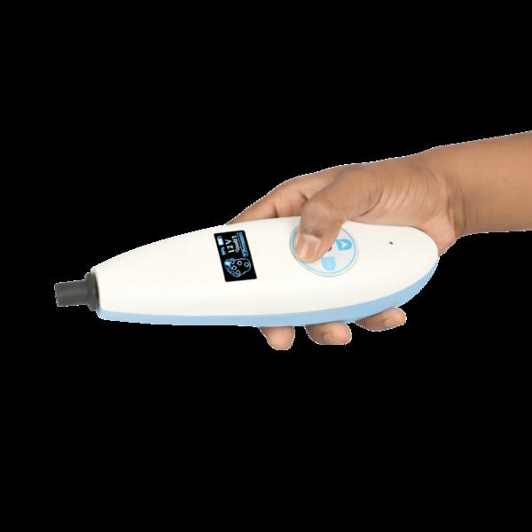
Skin Perfusion Pressure - SPP:
Skin perfusion pressure is an alternative technology to TCOM for assessing the perfusion status of skin or “skin capillary blood pressure.” Using the laser Doppler and pressure cuff in combination, provides the SPP measurement in mmHg. Normal perfusion for lower extremity SPP values is > 50 mmHg. A SPP measurement between 30 and 50 mmHg is diagnostic of PAD while an SPP measurement of < 30 mm Hg is diagnostic of severe PAD or CLI. The SPP is a clinically validated tool with a strong correlation to wound healing potential. The test is not affected by calcified leg arteries.
Our Product Range:
Handheld Vascular Doppler, Vascular Doppler Recorder, Automatic Vascular Doppler Recorder, Digital Biothesiometer, Podiascan, Foot Scanner, Pedography, Plantar Scan Systems, tcpO2, Monofilament 10gm, Neuropathy Therapy Stimulator, IR Light Theraphy, Podiatry chair and instruments are our major products.